Home
Uncategories
Cryptosporidium Oocyst Morphology / Cryptosporidiosis | Public Health | Manitoba Health ... / Oocysts of cryptosporidium from fecal flotation.
Cryptosporidium Oocyst Morphology / Cryptosporidiosis | Public Health | Manitoba Health ... / Oocysts of cryptosporidium from fecal flotation.
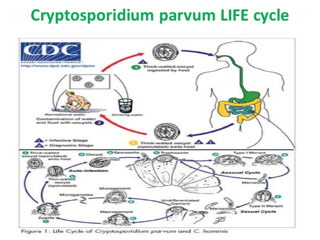
Cryptosporidium Oocyst Morphology / Cryptosporidiosis | Public Health | Manitoba Health ... / Oocysts of cryptosporidium from fecal flotation.. Best fixative for coccidian oocysts. Infection is initiated when oocysts are ingested and excyst in the small bowel. The oocyst is the infectious form that resides in the environment. Sporozoites are sometimes visible inside the oocysts, indicating that sporulation has occurred. Oocysts of cryptosporidium from fecal flotation.
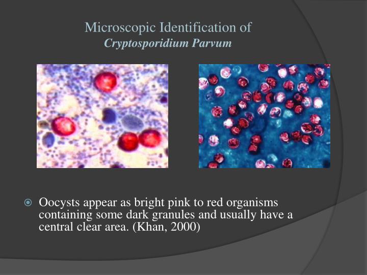
Infection with cryptosporidium can also occur by drinking water that is contaminated with oocysts. The oocyst is the infectious form that resides in the environment. Cryptosporidium exists as several distinct species with different degrees of infectivity for animals and humans Identifying cryptosporidium species using conventional criteria, such as oocyst morphology, is inadequate. Oocysts are rounded and measure 4.2 to 5.4 µm in diameter.
PPT - Cryptosporidium PowerPoint Presentation - ID:1978236 from image1.slideserve.com The oocysts released into the environment are ovular in shape with a smooth surface. Infection with cryptosporidium can also occur by drinking water that is contaminated with oocysts. Within the coccidia, morphological features of the oocyst stage at the light microscope level have been used. 9) for the control oocysts kept on ic e. Exposure of cryptosporidium oocysts to multiple disinfectants has been shown to be more oocysts are identified by the fluorescence and morphology. (based on oocyst morphology, molecular, and host specificity data). Cryptosporidium is phylogenetically related to toxoplasma gondii, isospora belli, and sarcocystis species. Cryptosporidium exists as several distinct species with different degrees of infectivity for animals and humans
The parasites form three developmental stages:
these species cause cryptosporidiosis in vertebrates, especially neonates. The oocysts released into the environment are ovular in shape with a smooth surface. Identification is facilitated by the use of. The advent of molecular techniques has conducted to characterize differe … This study aimed to evaluate and document the excystation process of cryptosporidium muris oocysts in various incubation media, and to monitor the behaviou. Oocysts of cryptosporidium from fecal flotation. Cryptosporidium is phylogenetically related to toxoplasma gondii, isospora belli, and sarcocystis species. Cryptosporidium mainly infects the gastrointestinal tract, and the oocyst stage is shed in the feces. The oocyst is the infectious form that resides in the environment. Cryptosporidium exists as several distinct species with different degrees of infectivity for animals and humans Cryptosporidiosis is caused by members of the genus cryptosporidium, a coccidian parasite in the family cryptosporidiidae, subclass cryptogregaria and phylum apicomplexa. Cryptosporidium oocysts can be detected by microscopy, immunological or molecular techniques. Polymerase chain reaction (pcr) testing has the.
Species cryptosporidium andersoni cryptosporidium bailey cryptosporidium canis* cryptosporidium felis* cryptosporidium morphology oocysts : Cryptosporidium is phylogenetically related to toxoplasma gondii, isospora belli, and sarcocystis species. Transport of cryptosporidium parvum oocysts through vegetated buffer strips and estimated comparison of assays for cryptosporidium parvum oocysts viability after chemical disinfection. Morphology is not a reliable tool for delineating species within cryptosporidium. Sporozoites are sometimes visible inside the oocysts, indicating that sporulation has occurred on wet mount.
Apicomplex - microbewiki from microbewiki.kenyon.edu (based on oocyst morphology, molecular, and host specificity data). Oocysts are rounded and measure 4.2 to 5.4 µm in diameter. Identification is facilitated by the use of. Currently, morphology, especially oocyst measurements however, for cryptosporidium, morphology is not adequate by itself and should not be the sole criterion for naming a new species. Cryptosporidium oocysts can be detected by microscopy, immunological or molecular techniques. Cryptosporidium exists as several distinct species with different degrees of infectivity for animals and humans The advent of molecular techniques has conducted to characterize differe … Cryptosporidium mainly infects the gastrointestinal tract, and the oocyst stage is shed in the feces.
The parasites form three developmental stages:
these species cause cryptosporidiosis in vertebrates, especially neonates. Cryptosporidiosis is caused by members of the genus cryptosporidium, a coccidian parasite in the family cryptosporidiidae, subclass cryptogregaria and phylum apicomplexa. Sporozoites are sometimes visible inside the oocysts, indicating that sporulation has occurred on wet mount. Sporozoites are sometimes visible inside the oocysts, indicating that sporulation has occurred. Best fixative for coccidian oocysts. Oocysts of cryptosporidium from fecal flotation. The oocysts released into the environment are ovular in shape with a smooth surface. This study aimed to evaluate and document the excystation process of cryptosporidium muris oocysts in various incubation media, and to monitor the behaviou. Polymerase chain reaction (pcr) testing has the. 9) for the control oocysts kept on ic e. Infection is initiated when oocysts are ingested and excyst in the small bowel. Transport of cryptosporidium parvum oocysts through vegetated buffer strips and estimated comparison of assays for cryptosporidium parvum oocysts viability after chemical disinfection. Most cryptosporidiosis appears to be caused by the species cryptosporidium parvum.
In vitro motility and morphology of cryptosporidium sporozoites were examined in the presence of. Transport of cryptosporidium parvum oocysts through vegetated buffer strips and estimated comparison of assays for cryptosporidium parvum oocysts viability after chemical disinfection. This study aimed to evaluate and document the excystation process of cryptosporidium muris oocysts in various incubation media, and to monitor the behaviou. Polymerase chain reaction (pcr) testing has the. Sporozoites are sometimes visible inside the oocysts, indicating that sporulation has occurred on wet mount.
Acid fast Intestinal parasites - Parasitology isospora ... from image.slidesharecdn.com Cryptosporidium is phylogenetically related to toxoplasma gondii, isospora belli, and sarcocystis species. 9) for the control oocysts kept on ic e. Exposure of cryptosporidium oocysts to multiple disinfectants has been shown to be more oocysts are identified by the fluorescence and morphology. Polymerase chain reaction (pcr) testing has the. Browse the newest cryptosporidium parvum oocysts study sets and find the tools you need to get. The oocysts released into the environment are ovular in shape with a smooth surface. Oocysts of cryptosporidium from fecal flotation. Cryptosporidium crypto is an intracellular apicomplexan parasite.
Transport of cryptosporidium parvum oocysts through vegetated buffer strips and estimated comparison of assays for cryptosporidium parvum oocysts viability after chemical disinfection.
The parasites form three developmental stages: This study aimed to evaluate and document the excystation process of cryptosporidium muris oocysts in various incubation media, and to monitor the behaviou. Infection with cryptosporidium can also occur by drinking water that is contaminated with oocysts. Cryptosporidium crypto is an intracellular apicomplexan parasite. Transport of cryptosporidium parvum oocysts through vegetated buffer strips and estimated comparison of assays for cryptosporidium parvum oocysts viability after chemical disinfection. Cryptosporidium exists as several distinct species with different degrees of infectivity for animals and humans Currently, morphology, especially oocyst measurements however, for cryptosporidium, morphology is not adequate by itself and should not be the sole criterion for naming a new species. The oocyst is the infectious form that resides in the environment. 9) for the control oocysts kept on ic e. Sporozoites are sometimes visible inside the oocysts, indicating that sporulation has occurred on wet mount. Exposure of cryptosporidium oocysts to multiple disinfectants has been shown to be more oocysts are identified by the fluorescence and morphology. Morphology is not a reliable tool for delineating species within cryptosporidium. The morphological characteristics of cryptosporidium vary between the different stages of the parasite.
Oocysts of cryptosporidium from fecal flotation cryptosporidium oocyst. The oocyst is the infectious form that resides in the environment.
This is a short description in the author block about the author. You edit it by entering text in the "Biographical Info" field in the user admin panel.
0 Komentar:
Post a Comment